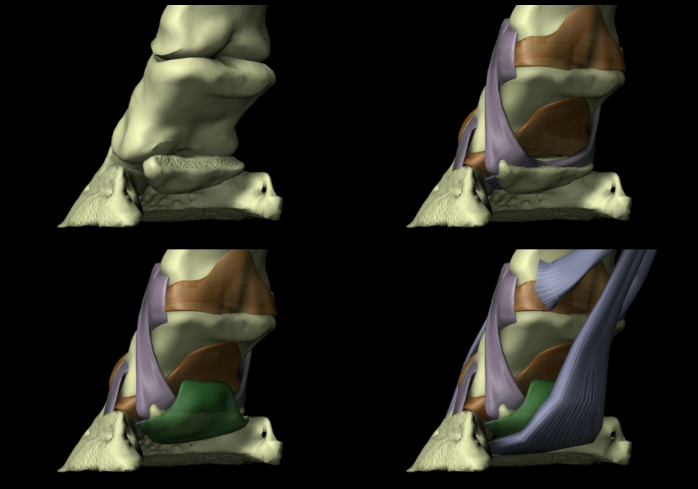
As established in Navicular Disease – Part 1: Background, navicular disease is damage to tendon, cartilage, and bone at the interface of the deep digital flexor tendon (DDFT or DFT) and the navicular bone as the consequence of heat generated from friction. The friction is the product of slow and/or fast vibration from improper (non-zero-coffin-joint-acceleration) landings, and the disease is the cumulative effect of the heat over a long period of time rather than the result of a singular incident.
As anyone who’s been around the horse world for any length of time undoubtedly knows, a diagnosis of “navicular” is incredibly common. Many veterinarians diagnose navicular syndrome or just plain “navicular” in situations where they see pain in the caudal (rearmost) portion of the hoof they can’t otherwise explain, and diagnose navicular disease whenever they see caudal hoof pain coupled with any sort of radiographic anomaly with the navicular bone.
In my experience, these diagnoses are wrong far more frequently than they’re right. Over the past 20-something years, I’ve examined many horses that have been diagnosed with some sort of “navicular” problem; yet, only 2 or 3 of those horses have had any evidence of what Dr. Rooney would’ve called “genuine” navicular disease. The rest have, in reality, been suffering from other issues – and, I might add, recovered from their lamenesses once the real causes of their problems were identified and properly treated. Just a few examples…
- I was asked to examine a horse the veterinarian had declared to be in pain due to either her right stifle or her right front navicular bone. She arrived at this diagnosis purely through observation and a flexion test on all four limbs – no physical examination, nerve blocks, or other diagnostic techniques were used. More disturbingly, she apparently didn’t realize that navicular issues would be very far down the list of possible diagnoses for this horse – a middle-aged Morgan broodmare with alleged symptoms in only one foot. As it turned out, the alleged “symptom of pain” ended up being nothing more than normal equine behavior, much to the relief of the owner!
- I received a call from a horse owner whose veterinarian had diagnosed “navicular,” and instructed the owner to have “special shoeing” used on the horse to “cure” the problem. After a year of following the veterinarian’s advice with no improvement in the horse, the owner contacted me through the university to see if anything could help his horse. Again – there was nothing about this horse that should’ve led to a diagnosis of “navicular;” she was only 6 or 7 years old, and had only been used for trail riding. A 30-second examination revealed that the horse had a very bad infection in the frogs of all four feet. The horse returned to complete soundness after treating the infections.
- One of the most glaring and nearly disastrous cases of misdiagnosis I’ve yet encountered has already been recounted in Toy Story. In that instance, a diagnosis of navicular disease by several veterinarians, including a so-called “hoof specialist,” nearly cost this horse his life. Once the real problem was diagnosed (White Line Disease) and treated, this horse went on to win a state championship!
Unfortunately, these are by no means isolated cases, but relating more of them here won’t serve any particular purpose. The above anecdotes are absolutely not meant to suggest that navicular disease isn’t real – it definitely is. But it’s certainly not the first or the second or the third thing horse owners or veterinarians should suspect when a horse presents with a lameness, and correctly diagnosing it, particularly in its early stages, entails a thorough understanding of its causes as well as what it is not. And so, when I speak of “genuine” navicular disease, I’m referring only to the condition resulting from actual DFT/navicular bone damage, and not the myriad other presentations of symptoms that end up labeled as “navicular” but really aren’t.
So how is genuine navicular disease diagnosed? Let’s start with the typical symptoms. In the beginning stages of genuine navicular disease, a horse:
- Will exhibit some degree of vague forelimb lameness affecting both limbs
- May alternately “point” his front feet
- May exhibit a foreshortened stride
Obviously, any number of conditions may account for these same symptoms, including mild laminitis and, especially, a horse that’s heel-sore from excessive concussion (much more on that later!). At this point in the diagnostic process, your veterinarian should be thoroughly palpating the limbs for any heat, swelling, and/or tenderness, as well as examining the hooves for signs of bruising, frog infection, and/or abscessing. Just bear in mind that although it’s unlikely (but not unheard of) to have simultaneous abscesses in both front feet, it could also be a combination of problems, such as an abscess in one foot and a pulled tendon in the other leg. Watching the horse move forward, backward, and turn is also very important to help rule out soft-tissue injuries higher up in the body, like sore shoulders or hips.
It’s imperative that any diagnostic work also include the horse’s history. Things like a recent change in hoof care providers or yesterday’s turnout in the mud after being stalled for a week can provide valuable insight into where to look – and, just as importantly, where not to look – for the potential source(s) of lameness.
Assuming other possibilities above the foot have now been eliminated and the horse’s symptoms are consistent with the preceding indications, the answers to the following questions will help include or eliminate genuine navicular disease from the list of diagnosis possibilities:
- How old is the horse?
- Does the horse have disproportionately-small feet for his body size?
- Is the horse shod?
- Does the horse have an obvious heel- or toe-first landing at the walk?
- Has the horse been extensively used for jumping, or on pavement or hard ground at speeds faster than a walk?
As mentioned in my examples above, this “equine profiling” process of evaluating risk factors will tend to “stack the deck” either against, or in favor of, a (correct) diagnosis of navicular disease. Since this condition is the result of repeated high-speed or high-tendon-travel-distance (as in jumping) heel- or toe-first landings, a young horse used only for flat work on soft ground is an extremely unlikely candidate for navicular disease; for example, a reining horse. On the other hand, an older horse with a lifetime of “corrective shoeing” that’s been used extensively for cross-country work, or a shod horse with an obvious heel-first landing and a history of extensive use pulling a cart on pavement, is much more likely to have genuine navicular disease.
If the horse’s physical characteristics and history still haven’t ruled out navicular disease, then your veterinarian may suggest nerve blocks as a “next step” in the diagnostic process. By injecting a small amount of a local anesthetic such as mepivacaine HCl into and around the palmar digital nerve, sensation in the hoof can be blocked. In horses with foot pain, the horse will generally “block sound,” or cease to show the lameness, regardless of the cause of the lameness. Because navicular disease is nearly always a bilateral condition i.e. one affecting both legs, the apparent lameness of a horse with genuine navicular disease will move to the opposite leg when either leg is nerve blocked. If it doesn’t, the cause of pain is very probably not navicular disease!
Note that radiographic evidence hasn’t been mentioned at all, and for a very good reason. According to Dr. Rooney –
The x-ray is of little or no use other than to muddle and confuse the picture in the early stages of navicular disease. It can be diagnostic, however, in advanced cases….The first true sign of navicular disease on x-ray is the osteophytes forming around the margins and radiolucent foci in the central area of the navicular bone (where the bone is being reabsorbed and replaced by connective tissue).
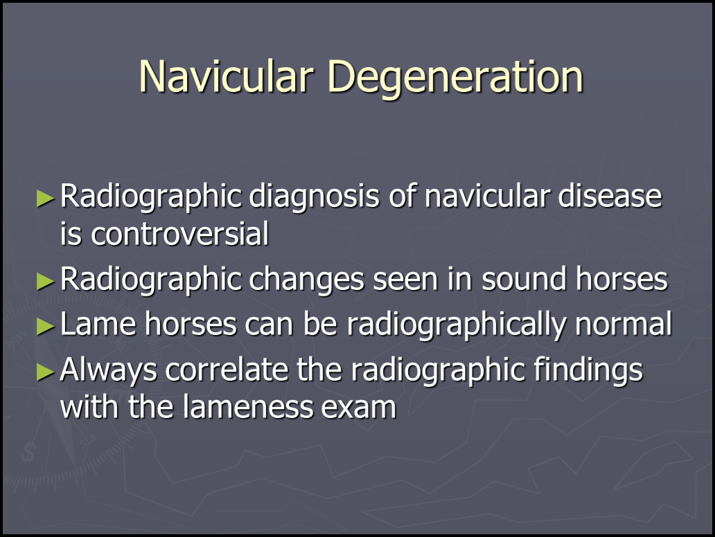
And Dr. Rooney isn’t the only one to recognize the potential problems with relying on radiographs to diagnose navicular disease. Take a look at this PowerPoint slide from Dr. Federica Morandi’s VM855 Veterinary Radiology class at the University of Tennessee –
So the “bottom line” on the use of radiographs for diagnosing navicular disease is this: if it’s an early case of navicular disease, x-rays will not give you a definitive answer either way. The only instance where a radiograph might be useful, then, would be to help differentiate between advanced navicular disease and some other pathology severely affecting both forelegs, such as two fractured coffin or navicular bones.
I also haven’t discussed the use of hoof testers in diagnosing navicular disease, for several reasons – most of which apply to using hoof testers in general. First of all, with sufficient force, a response can be elicited from nearly any horse. Second, since they aren’t calibrated, their use relies heavily on the ability of the person doing the testing to apply a consistent amount of pressure to the suspect and the “normal” hoof. And third, their use also depends on comparing the relative amount of force required to elicit a response on the suspect foot versus the “normal” foot, which, in the case of navicular disease, should be very similar as it’s a bilateral condition! So I think there are more accurate and reliable ways to determine whether or not a hoof is foot-sore.
On the other hand, one extremely useful diagnostic test in cases of suspected navicular disease that’s rarely done in the U.S. is the board test. I’m not certain why it’s so uncommon here; perhaps because it’s noninvasive and easily done using only a plank, veterinarians don’t feel they could justify charging enough for it! But according to Dr. Rooney, it’s a very simple way to eliminate DFT/navicular bone issues from the list of possibilities. If you’ve ever seen a flexion test, you’ve watched the veterinarian deliberately over-flex the coffin, pastern, and fetlock joints for some period, and then immediately walk the horse off and watch for lameness. A board test is essentially the same type of test, except we’re flexing the DFT/navicular bone interface. Here’s Dr. Rooney’s description of the board test in The Lame Horse (1998) –
Place a stout board on the ground in line with the horse and one front foot. Place the foot on the end of the board and lift the other end to about knee height and hold it. Eventually, the horse will take his foot off the board. If he puts the foot flat on the ground, the test is negative. If he immediately stands toe-first on the ground, it is positive and suggests navicular disease. The board test increases the pain because it increases the tension in the deep flexor tendon and the pressure exerted by the tendon on the surface of the navicular bone.
Note that this test, like others, will have false positives because horses can be heel-sore for a variety of reasons. But a negative test, even in one foot, will practically rule out navicular disease as a possible diagnosis, which is precisely why I like this simple, noninvasive test!
Probably the single most useful test for diagnosing genuine navicular disease, particularly early in the course of the disease, is magnetic resonance imaging (MRI), because both the soft-tissue (DFT) damage and the beginnings of damage to the cartilaginous surface of the navicular bone can be seen. Unfortunately, MRI facilities for horses are not (yet) very common, and the test is quite expensive. Even so, diagnosing navicular disease still requires that the veterinarian understand what navicular disease is and isn’t.
So, when trying to come up with an answer as to why a particular horse is lame, the possibility of navicular disease will almost certainly cross someone’s mind if the cause isn’t immediately obvious. Just keep in mind this devastating disease is actually much less common than many believe, and reaching a correct diagnosis in its early stages can be greatly helped by understanding why a horse’s physical characteristics and history either support or refute this diagnosis. And keep in mind that, as Dr. Rooney states, “no single test will permit diagnosis of navicular disease,” so if your veterinarian or hoof care provider is suggesting otherwise, or not asking the questions listed above, consider another opinion!
In the last installment in this series, we’ll discuss options for the horse who does, in fact, have genuine navicular disease.
More later!

