Snake oil poster

“Once begun this disease process is irrevocable and unremittingly destructive. There is no cure, no return to normal….It is no doubt true that “cures” of navicular disease with any form of treatment reflect an incorrect diagnosis. One does not cure bona fide navicular disease.”

– James R. Rooney, DVM

Difficult words to hear and accept, to be sure, from a man who was undoubtedly one of the few in a position to make such a statement. But before losing all hope for your “navicular” horse, please keep in mind the two very important points I made in Navicular Disease – Part 1: Background and Navicular Disease – Part 2: Diagnosis:

  1. Genuine navicular disease is damage to the deep flexor tendon and the attendant surface of the navicular bone caused by repeated heating of the tendon and bone from friction of the tendon moving across the bone, and,
  2. Genuine navicular disease is much less common than the very large number of (mis)diagnosed horses would lead us to believe.

Understanding and acknowledging both of these points is absolutely crucial to appreciating the existence of so many claims of “curing” navicular disease. According to Dr. Rooney, any such claim of a cure can only mean one thing: the horse in question never actually had navicular disease!

A moment’s thought will reveal why this surely must be true. While the DFT damage might, conceivably, heal if the damage to the navicular bone could somehow magically go away and not perpetuate the damage to the DFT, curing the damage to the articular surface of the navicular bone is no more likely in horses than it is in humans. Why would we see so many knee and hip replacements in people if joint damage could be undone with a dietary supplement or special shoes? Obviously, research into the treatment of various diseases is ongoing, but until someone demonstrates an effective, non-surgical approach for regenerating human cartilage and bone, I certainly wouldn’t expect to see anything that works for navicular disease in horses.

So, at least for the time being, if a horse truly has navicular disease, that damage must be considered permanent. And while it can certainly be managed to some extent, depending on the severity of the disease, it cannot be cured. So since we can’t undo what’s been done, our only viable options are to concentrate on slowing the disease’s progression and minimizing whatever pain is already present. And although I’m certainly not qualified to offer advice on pain management through drug therapy, I do want to briefly comment on one of the most commonly-prescribed drugs for “navicular”  horses: isoxsuprine.

Dr. Rooney pointed out in several of his publications that the most common competing hypothesis to his evidence-based belief that navicular disease is caused by mechanical problems is that navicular disease is, instead, a vascular (blood vessel) disease. In those same publications, he also made some very compelling arguments as to why the vascular hypothesis cannot be correct. Regardless, others’ belief that the disease has vascular origins explains why isoxsuprine – a vasodilator – is so frequently prescribed. After all, if the disease is caused by blood flow problems, it might seem logical to prescribe a drug that purports to increase blood flow. Unfortunately, as pointed out in “The Effect of Oral Isoxsuprine and Pentoxyfilline on Digital and Laminar Blood Flow in Healthy Horses” (Ingle-Fehr, J.E., and Baxter, G.M., Veterinary Surgery, 28 (1999): 154-160), isoxsuprine apparently doesn’t increase blood flow in the horse’s foot! Specifically:

No statistically significant increases in DBF (digital blood flow) or LP (laminar perfusion) were detected over the 10 day treatment period with either isoxsuprine or pentoxyfilline….Neither isoxsuprine nor pentoxyfilline increased blood flow to the digit or dorsal laminae in healthy (non-laminitic) horses.

Granted, because their primary concern was with the use of these drugs in the treatment of laminitis, their study was conducted on what they determined to be laminitis-free horses without regard to other possible foot pathologies. I suppose one might argue that perhaps the drug does, indeed, work on horses with compromised foot circulation but not on normal horses, but that strikes me as highly unlikely. The researchers went on to conjecture as to why isoxsuprine appears to make some laminitic (not “navicular”) horses more comfortable, and concluded that the drug must have a very mild analgesic (pain-relieving) effect unrelated to circulation, since isoxsuprine apparently doesn’t affect blood flow.

For the “navicular” horse owner, therefore, the implications of this study are quite clear: whether or not your veterinarian believes navicular disease is caused by, or related to, circulation problems, isoxsuprine has been demonstrated to have no effect on blood flow in the equine foot. Refer him/her to the aforementioned article if he/she doesn’t believe you. Why give your horse an expensive, unnecessary, and ineffective drug? And if your horse does need relief from pain, there are far more effective and less expensive drugs readily available.

Through understanding the true cause of navicular disease comes the answer to slowing its progression: since the disease is the consequence of repeated heating of the tendon and bone from friction of the tendon moving across the bone, we must minimize the friction and consequent heating. How? Well, let’s review the list of factors that affect friction and heating from Navicular Disease – Part 1: Background:

  1. The degree of front-to-back imbalance in the hoof
  2. The stiffness of the hoof
  3. The hardness of the terrain upon which the horse moves
  4. The speed at which the horse moves
  5. The duration of the horse’s movement
  6. The size of the navicular bone and deep-digital flexor tendon

Given that we’re now talking about real-world horses with less-than-optimal hoof care, we have to add yet another factor that affects the “navicular” horse:

0. The actual length of the hoof relative to its optimal length

When a hoof is properly trimmed, it will be at its shortest possible length without compromising its structural integrity or increasing its sensitivity to terrain variations. Any length in excess of this optimal length, whether from growth or from the addition of a shoe, will increase the amount of time required for the hoof to leave the ground (breakover time) during maximum DFT tension across the navicular bone. Although this probably doesn’t contribute to friction and heating, it does place more strain in the damaged area of the foot we’re trying to protect.

Be aware also that the common practice of using “special” shoes on these horses, such as rolled, rocker, or squared-off toes, has not been found to significantly shorten breakover time; only proper hoof length can minimize breakover time (Back, W., and Clayton, H., Equine Locomotion, (2001): 146 & 149). Note these photos of left front legs at the moment of maximum DFT tension, just before the heels leave the ground –
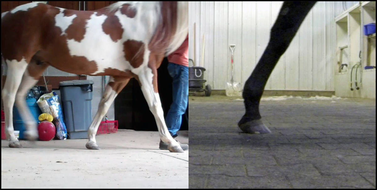
maximum-dft-strain

Adding length to these feet would only add to the time required for the hoof to leave the ground, thus prolonging the period of maximum strain across the navicular bone.

When managing the horse with navicular disease, we obviously have greater control over some of these factors than over others. For example, we cannot change the size of the horse’s bone and tendon, so number 6 can be eliminated from the onset. On the other hand, smart choices for the hardness of the terrain we ride on, coupled with how fast we ride and for how long we ride (numbers 3 through 5), can certainly minimize the amount of friction and heating of the bone and tendon. Remember: slower movement over softer terrain for shorter periods of time causes far less friction than faster movement over harder terrain for longer periods of time, just as less frequent jumps over shorter obstacles causes less friction than more frequent jumps over taller obstacles.

Far and away, though, our best opportunity for slowing the progression of the disease can be realized by minimizing numbers 0, 1, and 2 through proper hoof care. In concrete terms, that means:

  1. Properly trimming the horse so the hoof is optimally short and the coffin joint experiences minimal acceleration at landing i.e. a flat landing, and,
  2. Keeping the horse barefoot to maintain optimal hoof length, allow the foot to deform and absorb energy during initial ground contact and as it is loaded by the weight of the horse, and permit the most rapid breakover possible to minimize DFT strain across the navicular bone.

Unfortunately, these absolutely essential management measures are in diametric opposition to the advice of nearly every veterinarian and farrier. In fact, the most common advice given for the management of the “navicular” horse is the exact opposite of the above: use wedge shoes to elevate the heels and lessen the tension of the DFT across the navicular bone. While at first blush that course of action may seem logical, raising the heels of a horse makes only a small, temporary reduction in the tension of the DFT while simultaneously increasing the tension of the superficial digital flexor tendon, suspensory ligament, and extensor tendon. Much more problematic, however, especially for our “navicular” horse, is the increase in friction and consequent heating of the tendon and navicular bone that occurs with the resultant heel-first landing. In other words, elevating the heels (re)creates the very situation that caused the navicular disease in the first place!

The only situation where I could envision heel elevation as possibly being helpful to a horse with navicular disease would be if the horse were to be kept strictly on a hard, flat surface and limited to brief speeds of no greater than a walk. Think about it: if the horse were kept on a soft surface, the wedges would penetrate the surface with no net elevation of the heels, and if the horse were to move on the hard surface at any appreciable speed, the damage caused by the friction at the DFT/navicular bone interface would be greater than any benefit gained by the slight lessening in DFT tension through raising the heels.

So the first priority in managing the horse with navicular disease must be stopping the progression of the disease by not doing what’s probably been done, in terms of hoof care, for the majority of the horse’s life. Because of the widely-held, but incorrect, belief that horses are “supposed” to land heel-first (see The Myth of the Heel-First Landing series for more information on why this is incorrect), the product of many – I would even say “most” – hoof care providers’ efforts are feet that are incorrectly balanced in the front-to-back (A/P) direction and therefore experience a heel-first landing. As I’ve tried to make clear through this series of articles, long-term heel-first landings are the underlying cause of navicular disease, and the problems begin when horse owners fall into what I’ve termed “The ‘Navicular’ Trap” –
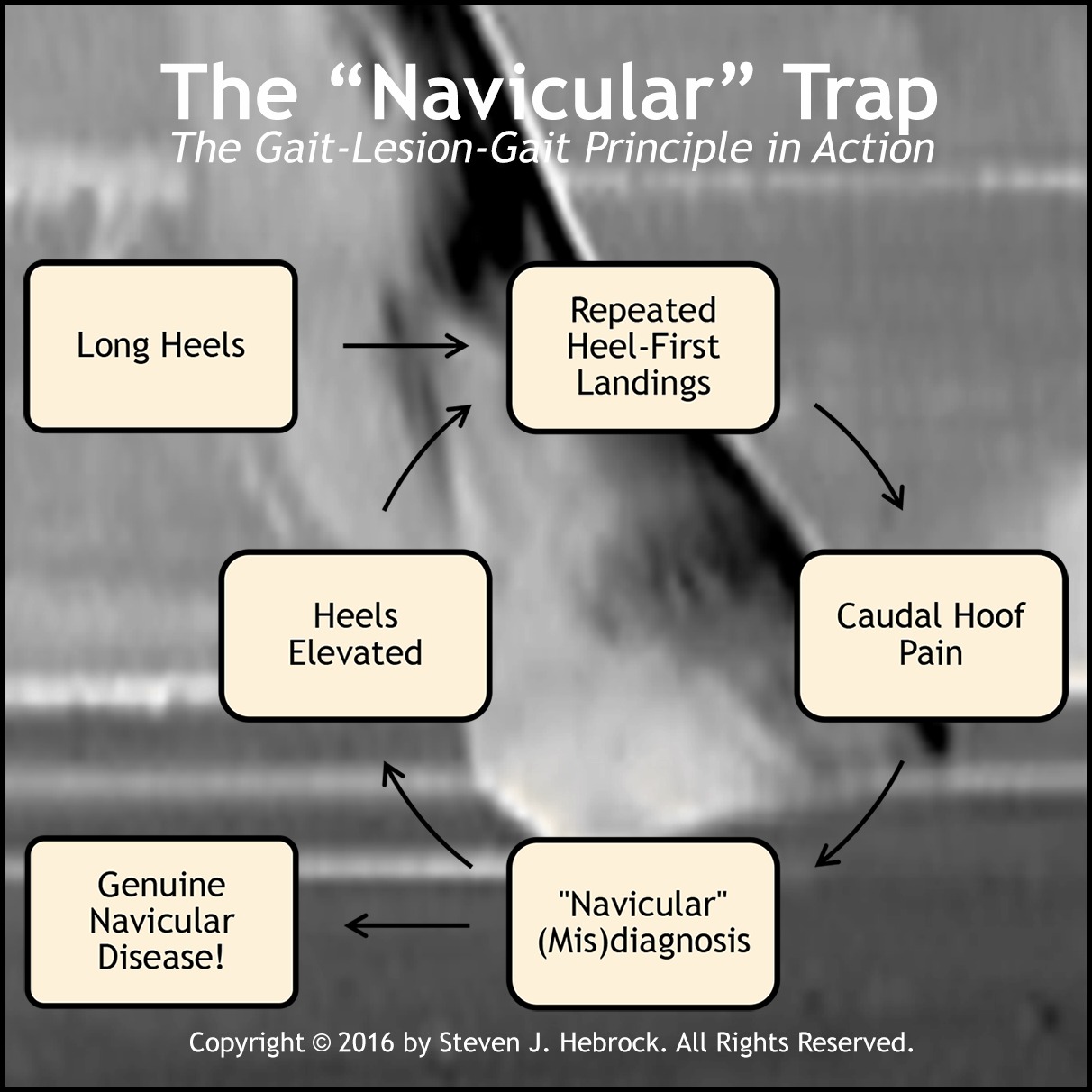
The Navicular Trap

Here’s how it usually plays out:

  1. The horse’s (front) heels are left too long by the hoof care provider
  2. As a result of working on harder ground, the horse becomes heel-sore from repetitive pounding
  3. The vet or hoof care provider diagnoses “navicular,” and raises the heels to allegedly reduce pain
  4. The horse becomes increasingly heel-sore because of increased pounding
  5. As a consequence of the repeated friction and attendant heating at the DFT/navicular bone interface, the horse ends up developing genuine navicular disease

This is a perfect illustration of the so-called gait-lesion-gait principle in action. As Dr. Rooney phrased it in his Biomechanics of Lameness in Horses –

The gait abnormality caused by a specific lesion is the gait abnormality which will cause the lesion.

What does that mean? Well, I interpret it like this: if a horse (or anything else) is forced to move in a manner that mimics the gait of a particular pathology, continued movement in that manner will eventually cause the very pathology the gait is indicative of! Specifically, if a horse’s heels are left too long for too long, he will become heel sore and his stride will be foreshortened (a temporary gait abnormality indicative of navicular disease). If his heels continue to be left too long – or worse, are further elevated with wedges – he may eventually develop navicular disease (the lesion), which will then cause permanent heel pain (and a permanent gait abnormality).

So preventing navicular disease and managing the horse with existing navicular disease are actually one and the same process: ensure the horse is experiencing minimal coffin-joint acceleration to the extent possible, using the guidelines above. By doing so, both the sound horse and the “navicular” horse will be moving with minimal resistance and (therefore) maximal efficiency, giving him the best chance possible at long-term comfort and soundness. And that’s the goal!

To reiterate the most important points of this series of articles:

  • True navicular disease is damage to the deep flexor tendon (DFT) and the attendant surface of the navicular bone.
  • Navicular disease is the result of repeated heating of the DFT and navicular bone surface caused by the friction resulting from non-zero-acceleration coffin-joint landings i.e. toe-first or heel-first landings.
  • While it definitely does exist, instances of true navicular disease are far less prevalent than commonly believed.
  • Diagnosing navicular disease cannot be done via radiographs unless the disease is already in its advanced stages, and instead is best diagnosed with MRI or less accurately with a combination of techniques including a thorough patient history.
  • True navicular disease cannot be cured.
  • The two most common treatments for navicular disease – isoxsuprine and wedge shoes – are ineffective (isoxsuprine) and cause further damage (heel wedges).
  • Effectively managing navicular disease and preventing navicular disease both depend on minimizing the underlying cause of the disease (friction) through proper hoof care, which means an optimally-short hoof experiencing a flat landing at the walk.

Above all, don’t lose hope if your horse is diagnosed with “navicular;” in my experience, odds are he doesn’t really have it! It’s much more probable he’s sore from excessive heel length, bad side-to-side balance (i.e. “corrective trimming”), sheared heels from radically different heel lengths, or an infection in his frog – all problems related to improper hoof care. Carefully consider his history and symptoms as well; your halter horse or brood mare isn’t a likely candidate for navicular issues, and a diagnosis of “navicular” in a single front foot or hind feet is probably not correct, either. Find someone who truly understands what proper hoof care is all about (admittedly challenging!), and allow him/her to help you rule out other far more likely possibilities.

In wrapping up this series, please allow me to make just one more point: I’m well aware that much of what I’ve presented – indeed, much of what I present on a variety of subjects, not just on navicular disease – flies in the face of popular thinking and advice. But what it absolutely doesn‘t fly in the face of is logic. If you’ll set aside your beliefs and carefully consider the evidence I’ve presented, I think you’ll agree…
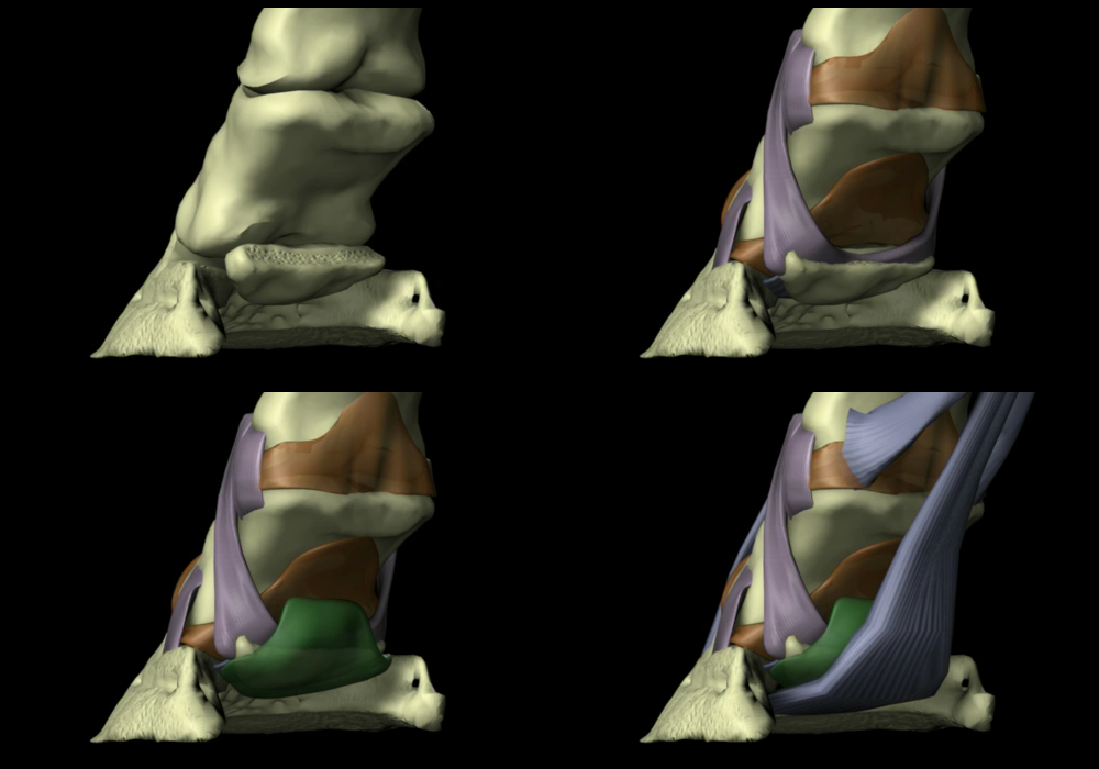
Glass Horse Navicular Composite
– still image captures from The Glass Horse: The Equine Distal Limb

As established in Navicular Disease – Part 1: Background, navicular disease is damage to tendon, cartilage, and bone at the interface of the deep digital flexor tendon (DDFT or DFT) and the navicular bone as the consequence of heat generated from friction. The friction is the product of slow and/or fast vibration from improper (non-zero-coffin-joint-acceleration) landings, and the disease is the cumulative effect of the heat over a long period of time rather than the result of a singular incident.

As anyone who’s been around the horse world for any length of time undoubtedly knows, a diagnosis of “navicular” is incredibly common. Many veterinarians diagnose navicular syndrome or just plain “navicular” in situations where they see pain in the caudal (rearmost) portion of the hoof they can’t otherwise explain, and diagnose navicular disease whenever they see caudal hoof pain coupled with any sort of radiographic anomaly with the navicular bone.

In my experience, these diagnoses are wrong far more frequently than they’re right. Over the past 20-something years, I’ve examined many horses that have been diagnosed with some sort of “navicular” problem; yet, only 2 or 3 of those horses have had any evidence of what Dr. Rooney would’ve called “genuine” navicular disease. The rest have, in reality, been suffering from other issues – and, I might add, recovered from their lamenesses once the real causes of their problems were identified and properly treated. Just a few examples…

  • I was asked to examine a horse the veterinarian had declared to be in pain due to either her right stifle or her right front navicular bone. She arrived at this diagnosis purely through observation and a flexion test on all four limbs – no physical examination, nerve blocks, or other diagnostic techniques were used. More disturbingly, she apparently didn’t realize that navicular issues would be very far down the list of possible diagnoses for this horse – a middle-aged Morgan broodmare with alleged symptoms in only one foot. As it turned out, the alleged “symptom of pain” ended up being nothing more than normal equine behavior, much to the relief of the owner!
  • I received a call from a horse owner whose veterinarian had diagnosed “navicular,” and instructed the owner to have “special shoeing” used on the horse to “cure” the problem. After a year of following the veterinarian’s advice with no improvement in the horse, the owner contacted me through the university to see if anything could help his horse. Again – there was nothing about this horse that should’ve led to a diagnosis of “navicular;” she was only 6 or 7 years old, and had only been used for trail riding. A 30-second examination revealed that the horse had a very bad infection in the frogs of all four feet. The horse returned to complete soundness after treating the infections.
  • One of the most glaring and nearly disastrous cases of misdiagnosis I’ve yet encountered has already been recounted in Toy Story. In that instance, a diagnosis of navicular disease by several veterinarians, including a so-called “hoof specialist,” nearly cost this horse his life. Once the real problem was diagnosed (White Line Disease) and treated, this horse went on to win a state championship!

Unfortunately, these are by no means isolated cases, but relating more of them here won’t serve any particular purpose. The above anecdotes are absolutely not meant to suggest that navicular disease isn’t real – it definitely is. But it’s certainly not the first or the second or the third thing horse owners or veterinarians should suspect when a horse presents with a lameness, and correctly diagnosing it, particularly in its early stages, entails a thorough understanding of its causes as well as what it is not. And so, when I speak of “genuine” navicular disease, I’m referring only to the condition resulting from actual DFT/navicular bone damage, and not the myriad other presentations of symptoms that end up labeled as “navicular” but really aren’t.

So how is genuine navicular disease diagnosed? Let’s start with the typical symptoms. In the beginning stages of genuine navicular disease, a horse:

  • Will exhibit some degree of vague forelimb lameness affecting both limbs
  • May alternately “point” his front feet
  • May exhibit a foreshortened stride

Obviously, any number of conditions may account for these same symptoms, including mild laminitis and, especially, a horse that’s heel-sore from excessive concussion (much more on that later!). At this point in the diagnostic process, your veterinarian should be thoroughly palpating the limbs for any heat, swelling, and/or tenderness, as well as examining the hooves for signs of bruising, frog infection, and/or abscessing. Just bear in mind that although it’s unlikely (but not unheard of) to have simultaneous abscesses in both front feet, it could also be a combination of problems, such as an abscess in one foot and a pulled tendon in the other leg. Watching the horse move forward, backward, and turn is also very important to help rule out soft-tissue injuries higher up in the body, like sore shoulders or hips.

It’s imperative that any diagnostic work also include the horse’s history. Things like a recent change in hoof care providers or yesterday’s turnout in the mud after being stalled for a week can provide valuable insight into where to look – and, just as importantly, where not to look – for the potential source(s) of lameness.

Assuming other possibilities above the foot have now been eliminated and the horse’s symptoms are consistent with the preceding indications, the answers to the following questions will help include or eliminate genuine navicular disease from the list of diagnosis possibilities:

  • How old is the horse?
  • Does the horse have disproportionately-small feet for his body size?
  • Is the horse shod?
  • Does the horse have an obvious heel- or toe-first landing at the walk?
  • Has the horse been extensively used for jumping, or on pavement or hard ground at speeds faster than a walk?

As mentioned in my examples above, this “equine profiling” process of evaluating risk factors will tend to “stack the deck” either against, or in favor of, a (correct) diagnosis of navicular disease. Since this condition is the result of repeated high-speed or high-tendon-travel-distance (as in jumping) heel- or toe-first landings, a young horse used only for flat work on soft ground is an extremely unlikely candidate for navicular disease; for example, a reining horse. On the other hand, an older horse with a lifetime of “corrective shoeing” that’s been used extensively for cross-country work, or a shod horse with an obvious heel-first landing and a history of extensive use pulling a cart on pavement, is much more likely to have genuine navicular disease.

If the horse’s physical characteristics and history still haven’t ruled out navicular disease, then your veterinarian may suggest nerve blocks as a “next step” in the diagnostic process. By injecting a small amount of a local anesthetic such as mepivacaine HCl into and around the palmar digital nerve, sensation in the hoof can be blocked. In horses with foot pain, the horse will generally “block sound,” or cease to show the lameness, regardless of the cause of the lameness. Because navicular disease is nearly always a bilateral condition i.e. one affecting both legs, the apparent lameness of a horse with genuine navicular disease will move to the opposite leg when either leg is nerve blocked. If it doesn’t, the cause of pain is very probably not navicular disease!

Note that radiographic evidence hasn’t been mentioned at all, and for a very good reason. According to Dr. Rooney –

The x-ray is of little or no use other than to muddle and confuse the picture in the early stages of navicular disease. It can be diagnostic, however, in advanced cases….The first true sign of navicular disease on x-ray is the osteophytes forming around the margins and radiolucent foci in the central area of the navicular bone (where the bone is being reabsorbed and replaced by connective tissue).

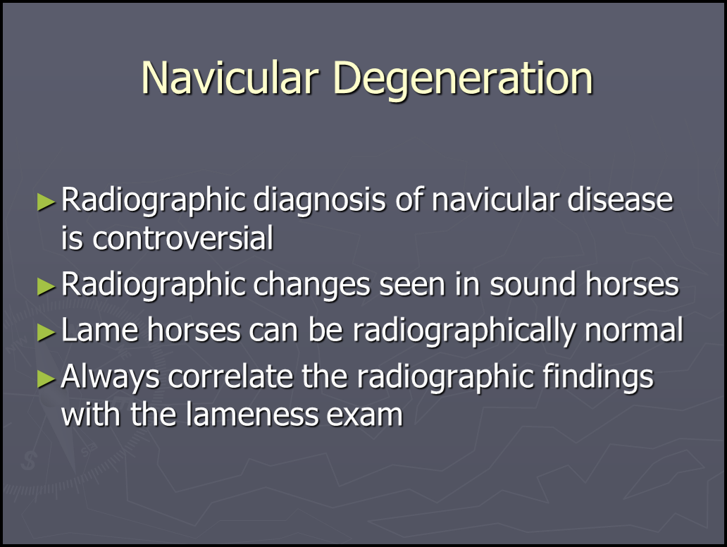
And Dr. Rooney isn’t the only one to recognize the potential problems with relying on radiographs to diagnose navicular disease. Take a look at this PowerPoint slide from Dr. Federica Morandi’s VM855 Veterinary Radiology class at the University of Tennessee –

Navicular Degeneration

So the “bottom line” on the use of radiographs for diagnosing navicular disease is this: if it’s an early case of navicular disease, x-rays will not give you a definitive answer either way. The only instance where a radiograph might be useful, then, would be to help differentiate between advanced navicular disease and some other pathology severely affecting both forelegs, such as two fractured coffin or navicular bones.

I also haven’t discussed the use of hoof testers in diagnosing navicular disease, for several reasons – most of which apply to using hoof testers in general. First of all, with sufficient force, a response can be elicited from nearly any horse. Second, since they aren’t calibrated, their use relies heavily on the ability of the person doing the testing to apply a consistent amount of pressure to the suspect and the “normal” hoof. And third, their use also depends on comparing the relative amount of force required to elicit a response on the suspect foot versus the “normal” foot, which, in the case of navicular disease, should be very similar as it’s a bilateral condition! So I think there are more accurate and reliable ways to determine whether or not a hoof is foot-sore.

On the other hand, one extremely useful diagnostic test in cases of suspected navicular disease that’s rarely done in the U.S. is the board test. I’m not certain why it’s so uncommon here; perhaps because it’s noninvasive and easily done using only a plank, veterinarians don’t feel they could justify charging enough for it! But according to Dr. Rooney, it’s a very simple way to eliminate DFT/navicular bone issues from the list of possibilities. If you’ve ever seen a flexion test, you’ve watched the veterinarian deliberately over-flex the coffin, pastern, and fetlock joints for some period, and then immediately walk the horse off and watch for lameness. A board test is essentially the same type of test, except we’re flexing the DFT/navicular bone interface. Here’s Dr. Rooney’s description of the board test in The Lame Horse (1998) –

Place a stout board on the ground in line with the horse and one front foot. Place the foot on the end of the board and lift the other end to about knee height and hold it. Eventually, the horse will take his foot off the board. If he puts the foot flat on the ground, the test is negative. If he immediately stands toe-first on the ground, it is positive and suggests navicular disease. The board test increases the pain because it increases the tension in the deep flexor tendon and the pressure exerted by the tendon on the surface of the navicular bone.

Note that this test, like others, will have false positives because horses can be heel-sore for a variety of reasons. But a negative test, even in one foot, will practically rule out navicular disease as a possible diagnosis, which is precisely why I like this simple, noninvasive test!

Probably the single most useful test for diagnosing genuine navicular disease, particularly early in the course of the disease, is magnetic resonance imaging (MRI), because both the soft-tissue (DFT) damage and the beginnings of damage to the cartilaginous surface of the navicular bone can be seen. Unfortunately, MRI facilities for horses are not (yet) very common, and the test is quite expensive. Even so, diagnosing navicular disease still requires that the veterinarian understand what navicular disease is and isn’t.

So, when trying to come up with an answer as to why a particular horse is lame, the possibility of navicular disease will almost certainly cross someone’s mind if the cause isn’t immediately obvious. Just keep in mind this devastating disease is actually much less common than many believe, and reaching a correct diagnosis in its early stages can be greatly helped by understanding why a horse’s physical characteristics and history either support or refute this diagnosis. And keep in mind that, as Dr. Rooney states, “no single test will permit diagnosis of navicular disease,” so if your veterinarian or hoof care provider is suggesting otherwise, or not asking the questions listed above, consider another opinion!

In the last installment in this series, we’ll discuss options for the horse who does, in fact, have genuine navicular disease.

More later!

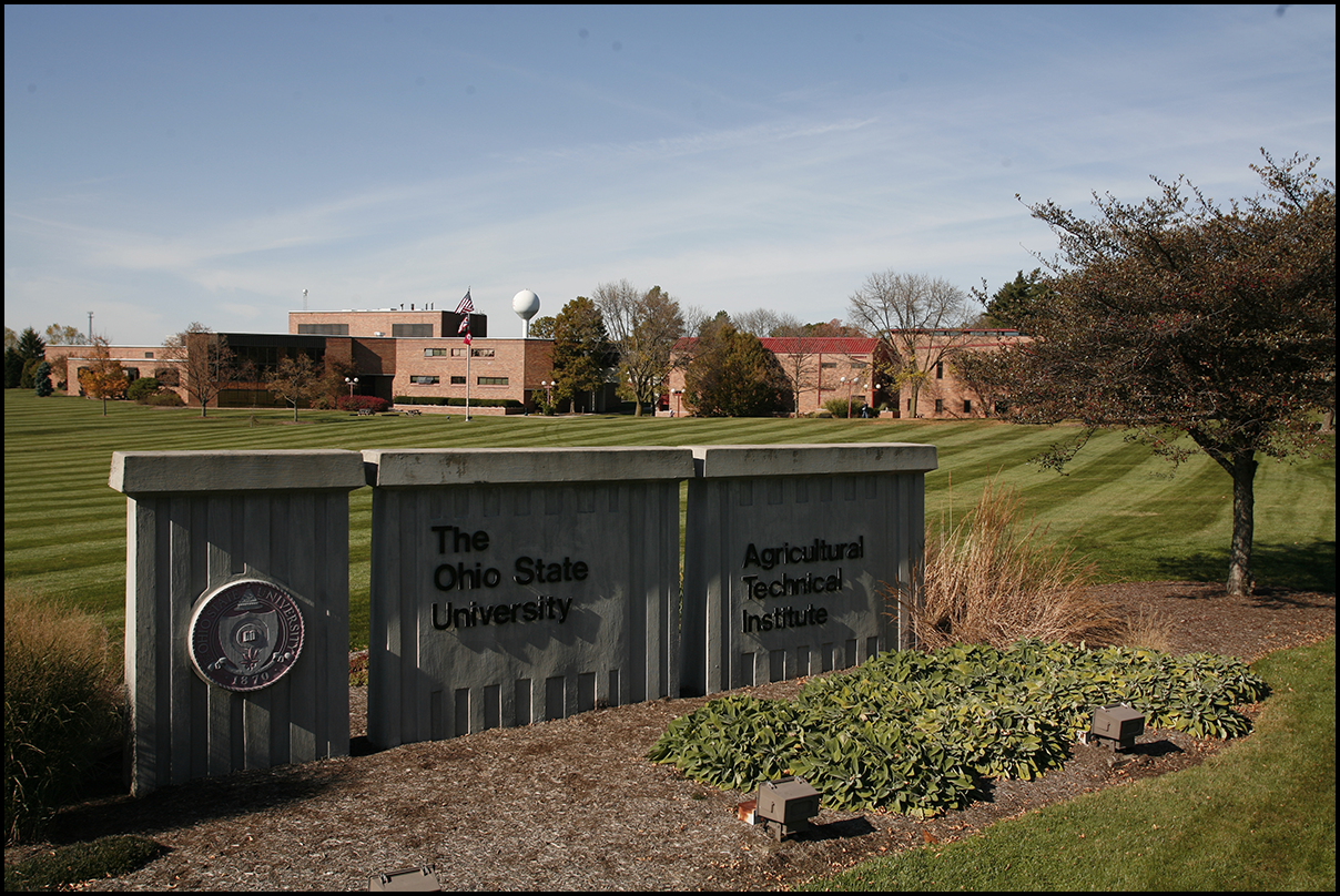
The Ohio State University ATI Campus

I’m very excited to announce that Liberated Horsemanship will be returning to our campus June 6th-10th for another Gateway Clinic! With an ever-increasing global interest in so-called “natural” hoof care on the part of both horse owners and hoof care providers, this clinic offers an unparalleled opportunity to learn proper hoof trimming theory and technique from industry-recognized experts.

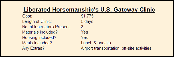
Why would you choose this particular training program over any of the others out there? Well, rather than point out what’s wrong with other hoof trimming training opportunities (which, to some extent, I’ve at least alluded to in previous articles), let’s take a look at some of the specifics of this clinic –

Liberated Horsemanship Clinic Information

While all of these factors undoubtedly figure into a person’s decision to choose a particular program, the most important issue in my mind has nothing directly to do with any of them. The deciding factor for me is the material being taught!

Unlike most, if not all, other training programs out there, Liberated Horsemanship’s teaching is absolutely consistent with the lessons learned about the determining factors of hoof form from the feral horses through the early efforts of natural hoof care pioneer Jaime Jackson; in other words, true natural hoof care. As such, this training program will equip the student to trim every hoof form successfully, regardless of size, breed, use, or pathology present.

The organization itself is unlike any other as well. Here are some statistics about just a few of its distinguishing characteristics –

Current number of program faculty:

Thirty (30)

Existing areas of specialization and research among the faculty:

Agronomy, architecture, biomechanics, booting, dentistry, endocrinology, engineering, ethology, hoof care, hoof pathology, laminitis, nutrition, parasitology, physiotherapy, veterinary medicine

Types and levels of certification currently offered:

Certified Hoof Care Practitioner, Certified Booting Specialist, Master Hoof Care Professional

Current instruction delivery methods:

Clinics, workshops, field instruction

For many people, two of the greatest benefits of this training program are the ongoing option of re-attending any previously-taken clinic or workshop at no additional cost (as many times as you want!), and the availability of the faculty as a support network. Quite a few of our students avail themselves of both of these; it’s fairly common to have students retake clinics 2 or 3 times, and receiving emailed or texted photos of unusual or problematic hooves along with questions is also a regular occurrence. And with such a diverse faculty, getting answers from recognized experts on difficult questions of not only hoof care, but on practically any equine-related issue, is relatively easy!

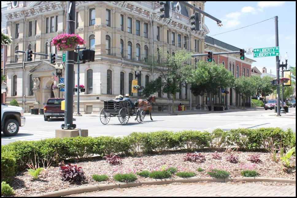
The learning environment for this clinic is also extremely pleasant. The agricultural branch of The Ohio State University, where the clinic is being held, is located in the small city of Wooster, Ohio, with a number of easily-accessible shops and restaurants on the edge of the largest settlement of Amish in the United States. Horse-drawn buggies and picturesque farms abound, and we’ll make at least one visit into Amish country to a very large farrier supply shop.

Downtown Wooster

As part of the clinic offering, there will also be a free, open-to-the-public event on Friday evening, June 10th featuring both Liberated Horsemanship and university faculty. Dr. Jessica Bedore will discuss the feeding issues associated with many older horses who have trouble maintaining body weight and condition. Master Hoof Care Professional Ann Corso will talk about hoof quality and its relationship to a horse’s overall health and happiness, and Dr. Bruce Nock will explain why obesity and insulin resistance – increasingly-common problems in our equine population – are not simply a matter of overfeeding. This promises to be an entertaining and informative evening, so regardless of whether or not you attend the entire clinic, please consider joining us for this special Friday night event!

Why Liberated Horsemanship YouTube Video

I strongly believe in this training program, and not just because I’m one of the faculty! As I explain in the brief YouTube video above, I truly believe this program offers unrivaled learning opportunities for everyone: prospective hoof care professionals, existing hoof care providers, and horse owners who wish to further their knowledge of proper horse management methods. Will you leave any clinic an “expert?” Of course not. But I guarantee you’ll walk away from this one with a whole lot more knowledge about horses and their hooves than you came in with, along with new friends beside you and an entire support network behind you.

Won’t you consider joining us?

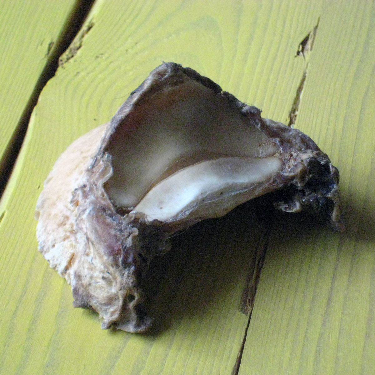
Photo of Coffin Bone with Navicular Bone

It’s a relatively small bone – about the size of a human thumb in the average horse. It bears no direct weight, doesn’t join any other bones together, and is rarely broken. And yet, it’s the alleged culprit in many, many cases of equine lameness. Without a doubt, no part of the equine hoof is as misunderstood, yet subject to incrimination for every bad step a horse takes, as the navicular bone. To quote Dr. James Rooney, the renowned equine pathologist –

Despite thousands, or perhaps millions, of words, navicular disease remains a subject of confusion and error.

There’s been a “method to my madness” in my past three articles (plus The Hoof Landings Tower of Babel, which wasn’t part of the original plan!) entitled The Myth of the Heel-First Landing. In addition to addressing the commonly-held misconception that horses are “designed” to land heel-first, the material was presented to help prepare the reader for coming to terms with the information I’ll be presenting on navicular disease. In this series of three articles, I’m going to attempt to shed some light on the navicular bone and hopefully allay some of the fears the horse owner experiences when he or she hears a diagnosis of “navicular” by the veterinarian. This particular installment will concentrate on an explanation of the role of the navicular bone in support and locomotion, and discuss how and why damage can occur. Part 2 will describe how navicular disease is diagnosed (and misdiagnosed), and the final article will focus on how to treat and/or manage the “navicular” horse.

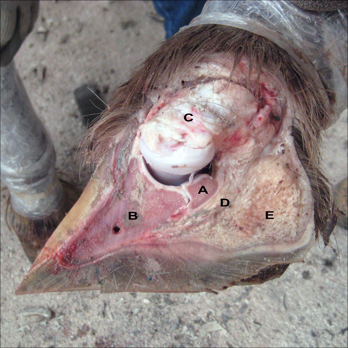
photo of cutaway hoof

As you can see in the photo at the beginning of this column, and in the above photo of a cutaway cadaver hoof, the navicular bone (A) lies right behind the coffin (pedal) bone (B) and forms part of the coffin joint along with the short pastern (C). You’ll also note the band of grey-white tissue running down the back/bottom surfaces of the bone that connects into the bottom center of the coffin bone. This is the deep digital flexor tendon (D), often abbreviated “DDFT” or simply “DFT,” which is responsible for both helping to stabilize the foot and flexing the foot towards the back of the horse. It also forms part of the “stay apparatus” of the horse – the horse’s suspension. Beneath the DFT is the largest structure in the foot – the fatty-fibrous mass of the digital cushion (E).

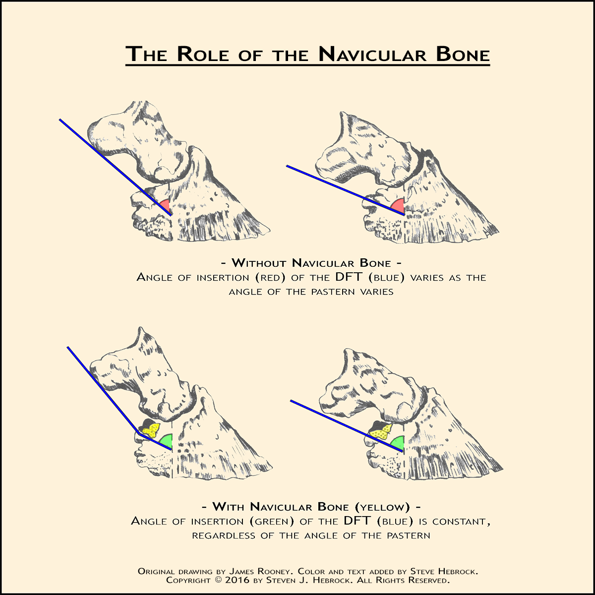
The navicular bone is one of three sesamoid bones found in the equine limb – the other two being the pair of small bones at the rear of the fetlock known as the proximal sesamoid bones. The purpose of a sesamoid bone is not to bear weight, but instead to change the direction of the pulling force being applied to a tendon. In this instance, the distal sesamoid bone, or navicular bone, ensures that the pulling force on the bottom of the coffin bone is always from the same direction, regardless of the angle of the pastern bones. Check out the following illustration –

The Role of the Navicular Bone

As you can see, without the navicular bone in place, the direction (and therefore the effective magnitude) of force exerted by the DFT would be highly dependent on the orientation of the long and short pastern bones with respect to the coffin bone. But with the navicular bone acting as a “pulley” to redirect the motion of the DFT, the force on the coffin bone will not change direction when the leg changes position.

So now we have a pulley (the navicular bone) with a rope (the DFT) running across it. Next, consider what Dr. Rooney wrote in Biomechanics of Lameness in Horses about what happens in the foot when the horse lands correctly (i.e. “flat”) –

As the hoof impacts, palmar flexion (rotation) of the coffin joint begins from zero acceleration. If the ground is hard, it will not yield under the hoof impact (will not absorb energy of impact), and the hoof may bounce, hit the ground, bounce again, etc. This bouncing, of course, would not be grossly obvious, but would involve oscillation of the coffin joint by a few millimeters. If the hoof as well as the ground were inelastic, the effect would, of course, be exaggerated. Neither ground nor hoof could yield sufficiently to quickly convert kinetic to potential energy. The energy would remain kinetic, oscillating back and forth between hoof and ground.

For the record: how do I know he’s describing a flat landing in the preceding quotation? The answer is simple: because he states “rotation of the coffin joint begins from zero acceleration.” And the only way that’s possible is if the hoof is contacting the ground flat. So even with a proper landing, there is some amount of vibration (oscillation) of the hoof, relative to the hardness of the landing surface and the inelasticity of the hoof capsule.

In contrast with the equine hoof, there is a considerable body of research on the effects of oscillations on humans’ hands and arms, although there is some dispute over various researchers’ conclusions – largely because differences in the types, frequencies, and durations of exposure used in various studies make analysis difficult. But the symptoms of hand-arm vibration syndrome (HAVS) among people who use various types of vibrating powers tools include destructive joint changes as well as vascular, neural, and muscular problems. In fact, you may be interested to know that it’s considered a serious enough health risk that the limits for vibration exposure for human workers are specified by the International Organization for Standardization (in ISO standard 5349-1)!

In the horse, when the coffin joint oscillates, the deep digital flexor tendon is moving very rapidly and repeatedly across the surface of the navicular bone. Why does that matter? Because when one surface moves rapidly over another surface, the friction between the two generates heating of the surfaces. Think warming cold hands, or starting a fire, by rubbing two things together – same idea. The more rapid and/or more sustained the motion, the higher the resulting temperature. Conversely, with slower and/or less-sustained movement, things don’t get nearly as hot. So, terrain and footwear being equal, a horse that’s moving at low speeds is experiencing less DFT/navicular bone friction, and therefore less heating, than the horse who is moving at higher speeds.

The problem gets much, much worse for the horse who has been trimmed or shod to contact the ground heel- or toe-first. In addition to the lesser amounts of vibration described above, he also experiences very rapid and more prolonged acceleration of the coffin joint at the instant of ground contact, called third-order acceleration, when the hoof “slaps” down as it comes under load. And the more pronounced the heel- or toe-first landing, the more rapid the acceleration and the greater the consequent heating. and landing heel-first or toe-first. That’s pure mechanics: the DFT of the horse who’s landing heel- or toe-first will be sliding farther, longer, and faster across the navicular bone than in a horse who’s landing correctly (flat), even at slower speeds. This is what Dr. Rooney had to say about third-order acceleration resulting from non-zero coffin joint acceleration at the moment of hoof contact i.e. a heel-first or toe-first landing –

In case it has not been said often enough, sudden changes of acceleration cause third-order acceleration, which is vibration: increased friction, turbulence, cavitation, heating, etc. The general case…is that third-order acceleration occurs between the DF and the navicular bone. Vibration, which is third-order acceleration, causes tendon degeneration and navicular arthrosis. The third-order acceleration, in turn, will occur because the coffin joint does not begin rotational movement from zero acceleration.

Sound familiar? It bears a striking resemblance to what’s been well-documented in humans experiencing HAVS, doesn’t it?

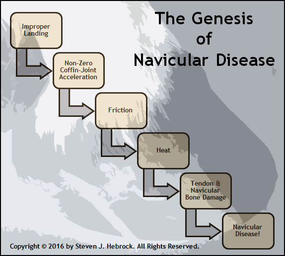
So in the simplest terms possible, folks, and in spite of what many people (including many veterinarians) believe, navicular disease is damage to the deep-digital flexor tendon and navicular bone caused by heat, which is caused by repeated heel-first or toe-first landings. In fact, Dr. Rooney, who spent a considerable amount of time researching this subject, reported that he was able to duplicate the physiology of navicular disease (bone and tendon damage) by using an external source of heat on cadaver limbs. Here, then, is an illustration of how and why navicular disease occurs –

The Genesis of Navicular Disease

This is also why feral horses don’t develop navicular disease. As I’ve explained in The Myth of the Heel-First Landing series, the feral horse’s hoof continuously wears itself such that, in general, the hoof (and therefore the coffin joint) does not experience rapid rotation as it makes contact with the ground i.e. it lands “flat.” And because the feral horse lands without the sudden “jerk” that occurs when many of our domestic horses make initial ground contact with the heels or toe and then “slap” the foot down as it comes under load (see the YouTube video accompanying The Hoof Landings Tower of Babel), the navicular bone in the feral horse doesn’t experience nearly as much rapid movement of the DFT across it with every step.

To summarize: whether or not a particular horse will develop navicular disease depends on not just one or two factors, but on a set of conditions that result in enough heating of the area of contact between DFT and navicular bone over a period of time to cause damage. It’s not a matter of any singular event causing the problem, but rather the cumulative effects of repeated “exposure” to vibration, similar to hearing loss or heavy-metal poisoning. All else being equal between horses, those factors include:

  • The degree of front-to-back imbalance in the hoof
    • The more pronounced the heel- or toe-first landing, the greater the distance the DFT travels across the navicular bone, resulting in higher/more-sustained temperatures.
  • The stiffness of the hoof
    • As the inelasticity of the unbalanced hoof increases, the less it can deform to mitigate the forces of impact, resulting in more vibration and consequent higher/more-sustained temperatures.
  • The hardness of the terrain upon which the horse moves
    • Like the stiffness of the hoof, the less the unbalanced hoof is able to penetrate the terrain at the instant of contact, the greater the vibration and resulting higher/more-sustained temperatures.
  • The speed at which the horse moves
    • The faster the horse is moving, the more frequent the coffin-joint rotation, and the greater the buildup of heat.
  • The duration of the horse’s movement
    • As with speed of movement, longer periods of movement of the unbalanced foot will result in higher/more-sustained temperatures.
  • The size of the navicular bone and deep-digital flexor tendon
    • The smaller the size of these structures, the less effectively they can dissipate heat buildup.

Looking at this list should make it apparent why “true” navicular disease (more on that subject later!) rarely occurs in horses that don’t either jump, or trot on paved roads. Those two activities, particularly when coupled with improper hoof care, experience the highest magnitude of (jumpers), and most prolonged (road horses), third-order acceleration of the coffin joint.

I also feel compelled to add that this explanation of the cause of navicular disease is by no means new. Dr. Rooney wrote The Biomechanics of Lameness in 1969! And while that particular book is admittedly quite technical and often difficult to read, both versions of his subsequent book The Lame Horse (1974 and 1998), which are intended more for the horse owner than the veterinarian, also describe this logical, evidence-based theory. Why, then, does there continue to be such a prevalent and profound misunderstanding of the cause of navicular disease among veterinarians, hoof care providers, and horse owners, with its consequent misdiagnoses and illogical “treatments” that cannot possibly succeed?

And that concludes Part 1 of this series! But after reading about the effects of vibration on humans (HAVS), I’m also very curious about whether or not some of the other consequences of vibration, like numbness, may occur in horses as well. Wouldn’t it be interesting to know if, over time, the unbalanced horse was actually losing sensation in his feet, and therefore allowing more damage to occur without his being aware of it?

One more thought to leave you with, which will undoubtedly be good news to many, but bad news for others –

In my experience, the overwhelming majority of horses diagnosed with navicular disease or navicular syndrome do not, in reality, have any issues with their navicular bone or deep-digital flexor tendon.

I’ll explain why I feel confident making that statement, as well as why so many horses are misdiagnosed, when I cover diagnosing navicular disease in the next article in this series.

Till next time…

Mary E. "Ginger" Hebrock
Mary E. “Ginger” Kuhn Hebrock 8/26/1934 – 9/14/2015

This Post has nothing directly to do with horses, so you may decide to ignore it. But, as I did for my father John Hebrock and for my best friend Gerald Bremseth, who both passed away in 2013, I must now do for my mother, who left this world on September 14th.

As I’ve often said about horse feet (ok, I did get horses in here!): it’s all about the balance. The same is true of nearly every aspect of life, and, in my life, my mother was in many respects the offsetting force that counterbalanced both my father’s strong drive towards perfection in everything he did, and his admitted lack of patience with anything and anyone that fell short of the mark.

Unfortunately, my father was also a very busy man, so projects around the house would often get started but not finished in any sort of a timely manner – simply because he didn’t have the time to do things exactly right, yet couldn’t bear to do things that weren’t perfect (I, too, suffer from this particular malady). My mother, on the other hand, was far more concerned with getting things done, whether they were perfect or not. A great example of this “difference in styles” could be seen in the main bathroom of the home I grew up in.

In 1962, my parents decided to purchase an unfinished house – the framing was completed, but nothing else – to finish themselves, which involved many, many trips to various lumberyards and supply houses with a trailer in tow. Because the bathroom was fairly narrow, my father spent a great deal of time and effort building a curved vanity with a matching cabinet door so one wouldn’t immediately walk into a sharp corner upon entering the bathroom. He spent hours on that cabinet, trying a wide assortment of construction techniques to achieve a door with an absolutely perfect arc. And the result was, indeed, perfect, and the rest of the bathroom came together quite nicely.

Except for the shelves and molding in the closet. Somehow, finishing them ended up a very low priority for my father, who had moved on to other projects once the rest of the bathroom was in hand. But not for my mother; she wanted the entire bathroom done, so although she’d frequently pester him about finishing, he never managed to return to it. So she finally took matters into her own hands, and cut the remaining pieces of lumber, nailed them up, and painted them. All blunt cuts, mind you – no miters! But it was done. And although my father was furious about her less-than-perfect work, that bathroom was still the same when the house was sold last year – 52 years later!

To be fair: she, too, was a perfectionist when it came to the things she was passionate about. Unlike his largely technical undertakings, my mother leaned heavily towards pursuits involving compassion for those in need. Trained as a registered nurse, she spent her life helping others: working in the pediatric ward of the local hospital, managing the needs of mentally and physically challenged children and adults in a county-run facility, caring for patients during ambulance transport, and other humanitarian efforts. It even extended to more “public” public service: she was the first female Village Trustee and Deputy Mayor of our small town in western NY.

And she had a great sense of humor! When we were kids, our grandmother (her mother) would often sew shirts as birthday or Christmas gifts for my brother and me, which we really enjoyed and appreciated. But shortly before one particular Christmas, my mother very solemnly sat us down to explain that our grandmother had also decided to give us bow ties with our new shirts, which my mother knew we intensely disliked. “Just pretend to be surprised and pleased,” she said. “You don’t want to hurt her feelings, after all.” Over the next days, we worried a lot about those bow ties and how we were going to manage to fool my grandmother into believing we were happy about them. And so, that Christmas morning, when our grandmother handed us the packages containing the shirts and bow ties, we both steeled ourselves to act pleased with the new additions to our wardrobe. And when we opened the boxes, we found our new shirts sporting bow ties…made out of $20 bills! They both had a terrific laugh over that!

But for many, one of her most memorable characteristics was her bright blue eyes, which often seemed to sparkle as if she were enjoying the most pleasant experience in the world that no one else was privy to. I remember seeing them shine many times in my younger days, especially when one of us accomplished something special like earning a good grade or playing an especially good concert. Indeed, her pride in her family and our achievements was always obvious, and shared with anyone who would listen.

Tragically, my mother was robbed of much of her vitality on a dark road one night in December of 1984, when my parents’ car struck the unlit trailer of a log truck a few miles from their home. Although both of them sustained multiple serious injuries in that crash, my mother suffered a head trauma that left her forever physically and emotionally impaired, and hastened the onset of dementia. A stroke in 2008 made things considerably worse, and necessitated her remaining in a nursing facility for the remainder of her life.

Still, she did what she could. Just a couple of years ago, one of the workers in the nursing home mentioned that my mother had been helping her study for her nursing exams, quizzing her on symptoms and treatments for cardiac disease. I wasn’t surprised by the news, because that was my mother – always helping others.

As anyone well-acquainted with my mother certainly knows, there’s a great deal more I could say about her. But this will have to suffice for now. Without a doubt, she touched the lives of many, many people in ways both acknowledged and unknown; as one of her caregivers of the last few years wrote to me, “I’ll miss your mother. She was a remarkable person.” And, along the way, my mother showed me over and over again that having compassion for others, and helping them to the best of one’s abilities, is an honorable and right and rewarding way to live.

I’m eternally grateful for what you so freely gave, mom. I will always miss you, but I’m glad you’ve finally found the peace you so richly deserve.

You were, indeed, a remarkable person…